Group Equivariant Diffusion for Anomaly Detection in Computational Cytology

Swarnadip Chatterjee, Ssharvien Kumar Sivakumar, Anirban Mukhopadhyay
MICCAI 2026
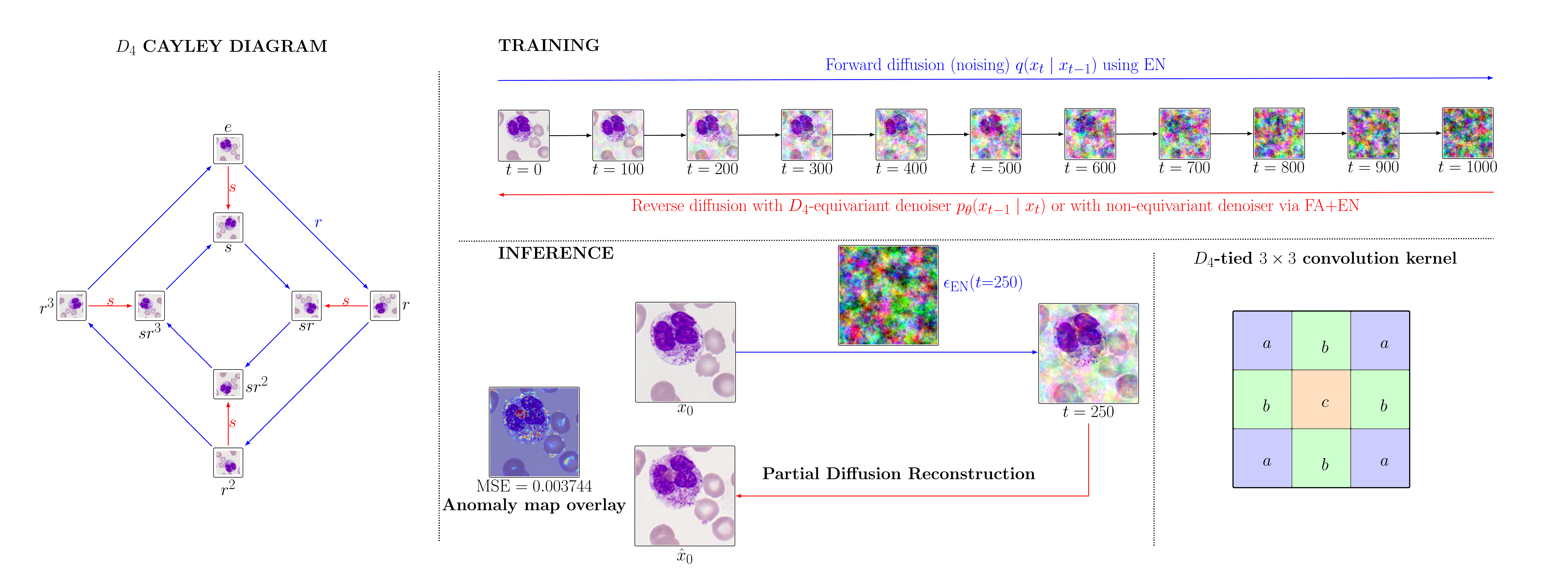
Overview of the D4-equivariant diffusion framework

D4diffCyto enforces rotation and reflection symmetry in diffusion-based anomaly detection for computational cytology.

Highlights

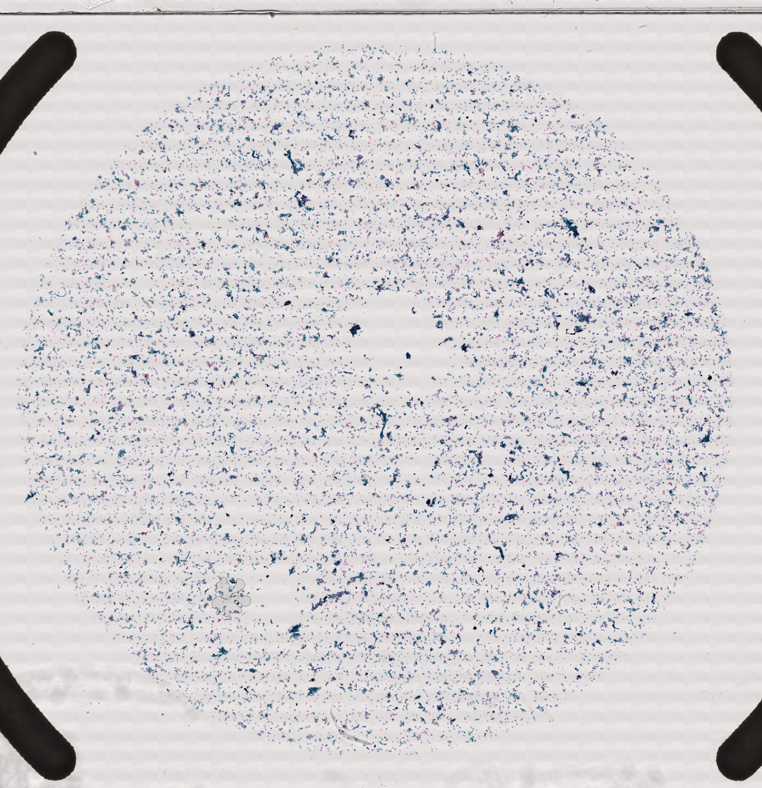
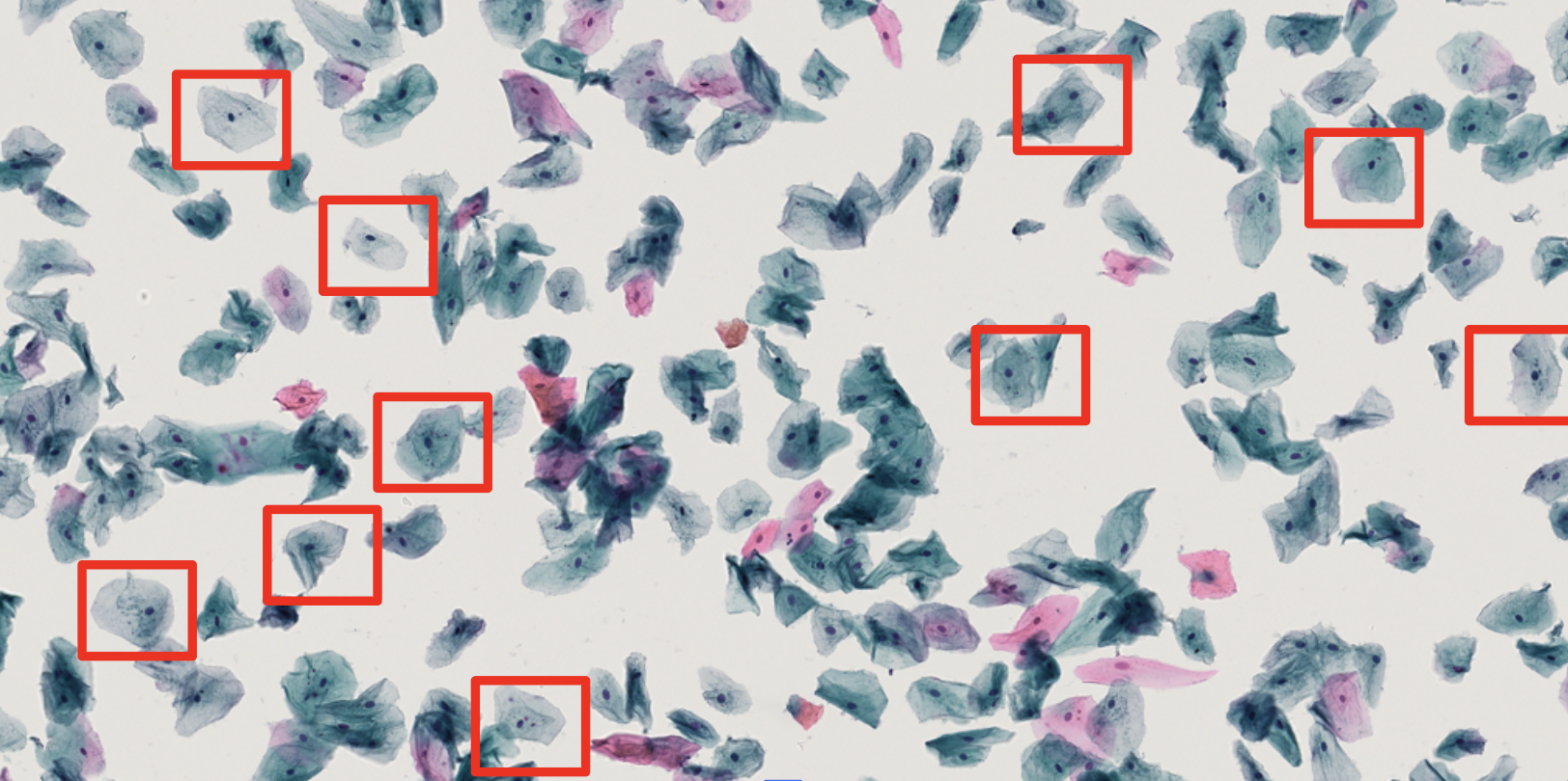
  • Computational cytology is challenging because abnormal cells are rare, heterogeneous, and expensive to annotate.
  • Diffusion anomaly detectors can be trained on normal slide-negative patches and then used to flag abnormal cells at test time.
  • A key problem is that standard diffusion models may produce different reconstructions and anomaly scores for rotated or flipped versions of the same cell patch.
  • D4diffCyto addresses this by enforcing dihedral symmetry, covering rotations and reflections of single-cell patches.
  • We study two complementary routes to D4-consistent anomaly scoring: inference-time symmetry enforcement using Frame Averaging and Equivariant Noise, and architectural symmetry enforcement using a D4-equivariant U-Net.
  • The method is evaluated on two public cytology datasets: MLL bone marrow cytomorphology and AML-LMU peripheral blood cytomorphology.
  • On MLL, FA+EN improves top-400 abnormal-cell retrieval from 57 to 78 compared with vanilla AnoDDPM.
  • On AML-LMU, the D4-equivariant U-Net with attention achieves the best AUC and top-400 retrieval among the evaluated diffusion variants.
  • D4-aware diffusion reduces anomaly-score variation across rotations and flips, making top-K retrieval more stable for realistic cytology screening workflows.

Motivation

Diffusion-based anomaly detection is attractive for cytology because it can be trained using only normal cells. A model learns a normal-cell appearance prior, and at test time an abnormal query patch is partially diffused and reconstructed toward the normal distribution. The residual between the input and the reconstruction becomes the anomaly signal.

However, single-cell cytology is different from organ-level medical imaging. In brain MRI, retinal OCT, or chest X-ray, anatomy often has a fixed orientation and lesions are spatially tied to that anatomy. In cytology, a centered cell patch can be rotated or flipped without changing its diagnostic meaning. A malignant or abnormal cell remains abnormal under the eight rotations and reflections of the square image grid.

This creates a structural mismatch: a standard diffusion model may treat rotated or flipped versions of the same cell as different inputs, leading to different pseudo-healthy reconstructions and different anomaly scores. Under extreme class imbalance, even small score changes can alter the top-K ranking and affect which cells are surfaced for expert review.

Rare abnormal cells Normal-only training Partial diffusion reconstruction D4 symmetry Stable top-K retrieval

From AnoDDPM to D4diffCyto

The starting point is AnoDDPM-style anomaly detection. Instead of generating a sample from pure noise, the test image is noised only up to an intermediate diffusion step and then denoised back. This partial diffusion reconstruction is useful because the model is trained on normal data, so the reverse process tends to reconstruct a pseudo-normal version of the query. The anomaly map is obtained from the reconstruction residual.

Input patch x0 → partial noising to xt* → reverse diffusion reconstruction x̂0
Anomaly map: |x0 − x̂0|
Anomaly score: mean squared reconstruction error

D4diffCyto keeps this partial-reconstruction principle but adapts it to cell-centric cytology. The key modification is that reconstruction should be consistent under the D4 group: rotations by 0°, 90°, 180°, 270° and their reflected counterparts. The goal is not only to obtain a good anomaly score, but to make the score stable under diagnostically irrelevant transformations.

What are FA and EN?

Frame Averaging (FA)

Frame Averaging applies the model to all transformed views of the same input and then maps the outputs back to the original orientation before averaging. For D4diffCyto, the frame is the D4 orbit of a cell patch: all rotations and flips of the same image.

Intuitively, FA asks the model: “What anomaly score would you give if the same cell appeared in every allowed orientation?” Averaging over these answers suppresses orientation-specific fluctuations and produces a more symmetry-consistent score.

FA score ≈ average over g in D4 of Score(g · x)

Equivariant Noise (EN)

Diffusion sampling is stochastic. Even if the denoiser is equivariant, independently sampled noise can make the trajectory for a rotated cell differ from the rotated trajectory of the original cell. EN fixes this by tying the random noise across transformed views.

For each D4 transform g, D4diffCyto uses the transformed version of the same base noise. Thus, the forward and reverse trajectories share the same underlying randomness up to the group action.

Base noise: ε
Noise for transformed view g · x: εg = g · ε

FA and EN play complementary roles. FA symmetrizes the outputs over the D4 orbit, while EN makes the stochastic diffusion trajectory itself consistent across transformed views. In D4diffCyto, the two are combined for inference-time D4 enforcement with a standard U-Net.

How FA+EN is adapted in D4diffCyto

In generic structure-preserving diffusion, FA and EN are used to make diffusion outputs respect a chosen transformation group. D4diffCyto adapts this idea to the specific operational problem of cytology screening: the system should rank abnormal cells consistently even when a cell appears rotated or flipped.

For inference-time D4 enforcement, a standard U-Net is first trained using the usual diffusion objective. No special group-equivariant architecture is required during training. At test time, the input patch is transformed by each element of D4. The same base noising variables are transformed accordingly, partial diffusion reconstruction is performed for each transformed view, residuals are mapped back to the original orientation, and scores are averaged.

  1. Start with a query cell patch x0.
  2. Generate the D4 orbit: rotated and flipped versions of x0.
  3. Use equivariant noise so each transformed view receives the correspondingly transformed stochastic noise.
  4. Run partial diffusion reconstruction for each view.
  5. Map anomaly maps back to the original orientation.
  6. Average residual maps and scalar scores over the orbit.
  7. Use the final averaged score for top-K abnormal-cell ranking.

This gives a simple way to enforce symmetry at inference time while retaining the ordinary U-Net training pipeline. It is especially useful when one wants to add symmetry consistency to a trained diffusion anomaly detector without redesigning the entire model architecture.

Abstract

Computational cytology on whole-slide images is challenging because malignant cells are rare, heterogeneous, and annotated slides are scarce. Anomaly detection frameworks can be trained on normal slide-negative patches and then applied at test time to flag abnormal patches in held-out slides. Most unsupervised anomaly detection approaches, including generative ones, are tuned to organ-level imaging and require large curated datasets. In cytology, the signal is cell-centric: rotating or flipping a single-cell patch does not change its diagnostic class, yet standard diffusion models treat transformed views as distinct inputs, leading to transformation-dependent reconstructions and unstable anomaly scores.

We propose a D4-equivariant diffusion framework that enforces rotation and reflection symmetry both architecturally, via a D4-equivariant U-Net, and at inference, via equivariant noise coupling and frame averaging. This alignment with biological invariance yields transformation-consistent pseudo-healthy reconstructions and more stable anomaly ranking under symmetry. On two publicly available cytology datasets of bone marrow and peripheral blood smears, our D4-equivariant diffusion models achieve higher AUC and retrieve more abnormal cells in the top-K predictions than non-equivariant generative baselines, a deep one-class baseline, and a multiple instance learning based method, while substantially reducing score variance across rotations and flips.
Method Overview

1. Normal-only training

The diffusion model is trained only on normal cytology patches. This is well suited to cytology, where collecting exhaustive abnormal-cell annotations is difficult, but normal or slide-negative examples are more accessible.

2. Partial reconstruction

At test time, the query patch is noised to an intermediate timestep and denoised back. The reconstruction is interpreted as a pseudo-normal version of the input, and the residual is used as the anomaly signal.

3. Top-K retrieval

Instead of only asking whether a whole slide is positive, the method ranks individual cell patches. This is important for realistic cytology workflows where only a small number of candidate cells may be inspected.

Inference-time D4 route: FA+EN

A standard U-Net is trained as usual. During inference, D4 consistency is imposed by applying rotations and flips to the input, coupling their diffusion noise through EN, reconstructing each transformed view, and averaging the resulting scores and residual maps using FA.

Architectural D4 route: D4 U-Net

The denoiser itself is made D4-equivariant using group-tied convolutional filters. This reduces orientation-specific variability inside the model and encourages the denoising function to respect rotations and reflections by design.

D4 U-Net with attention

The attention-augmented variant extends the D4-equivariant backbone with an attention block, allowing the model to capture richer spatial interactions while still preserving the intended group-aware structure.

These two routes are complementary. FA+EN is an inference-time mechanism that can be applied to a standard denoiser, while the D4-equivariant U-Net builds the symmetry into the architecture itself. Both are designed to reduce nuisance orientation variance and improve the reliability of abnormal-cell ranking.

Main Quantitative Results

Method MLL AML-LMU
AUC TP400 TP200 TP100 AUC TP400 TP200 TP100
Baselines
ItS2CLR 0.521107 27 20 19 0.360515 92 38 13
Deep SVDD 0.613331 49 35 21 0.514742 227 122 56
f-AnoGAN 0.594601 37 24 9 0.538857 273 131 57
THOR Gaussian 0.540851 24 14 7 0.582212 247 137 68
THOR Simplex 0.404264 8 3 1 0.544415 207 104 58
BerDiff 0.550799 44 32 26 0.708558 313 157 64
AnoDDPM 0.632846 57 30 18 0.694320 311 162 83
D4-equivariant diffusion variants
FA only 0.612543 46 32 19 0.680111 277 144 72
FA+EN 0.684121 78 53 34 0.678373 285 149 75
D4 conv. 0.675404 74 53 27 0.699635 315 159 86
D4 conv. + attn. 0.679251 67 44 33 0.720098 325 162 81

Patch-level anomaly detection results on MLL and AML-LMU. TP400, TP200, and TP100 denote the number of abnormal cells retrieved among the top-K most anomalous patches.

Qualitative Results

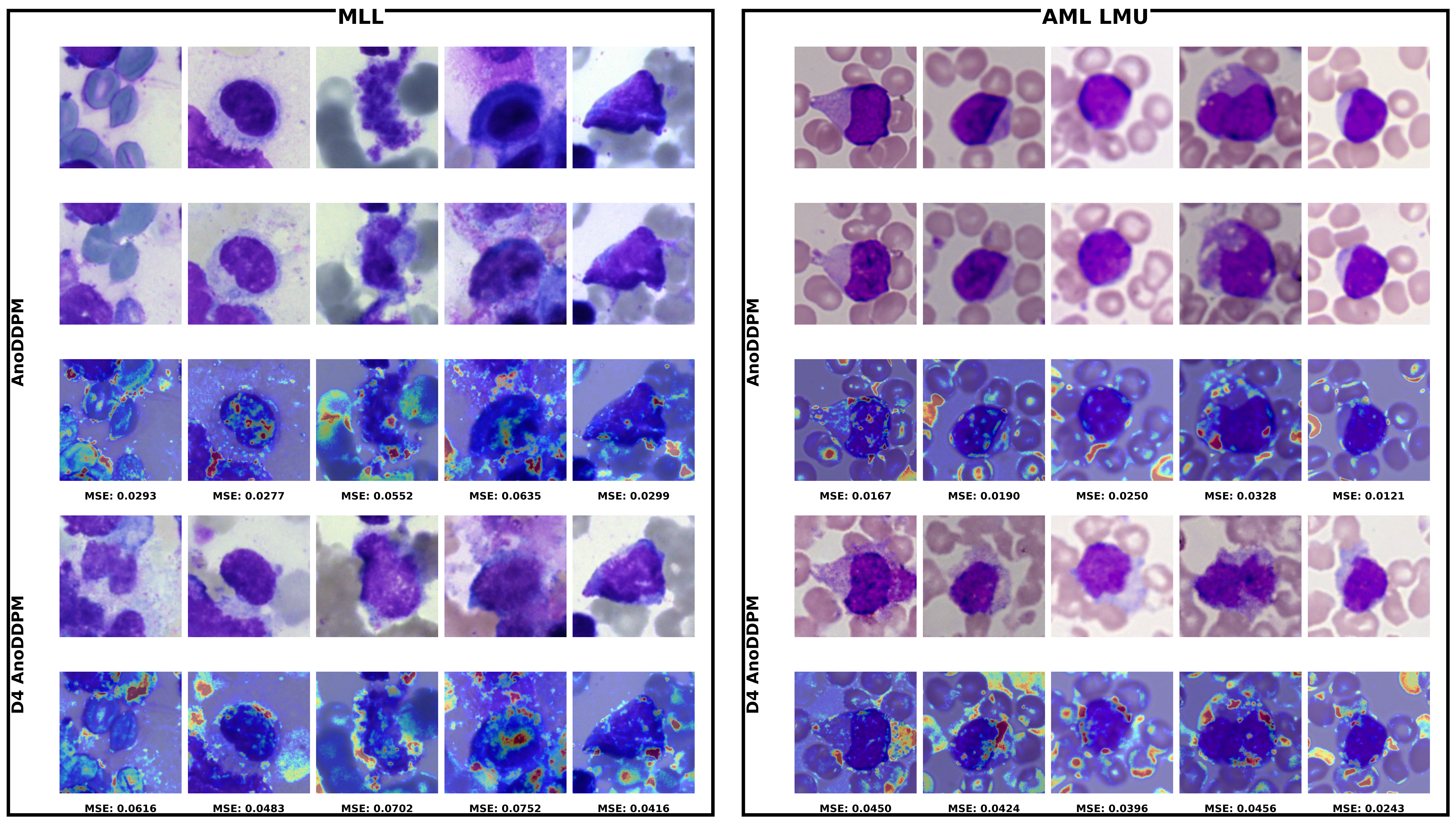
Qualitative comparison between AnoDDPM and D4-equivariant AnoDDPM

Qualitative comparison of vanilla AnoDDPM and D4-equivariant AnoDDPM on MLL bone marrow and AML-LMU peripheral blood cytology patches. Each panel shows inputs, reconstructions, and anomaly-map overlays with MSE scores.

BibTeX

@inproceedings{chatterjee2026d4diffcyto,

title     = {Group Equivariant Diffusion for Anomaly Detection in Computational Cytology},
author    = {Chatterjee, Swarnadip and Sivakumar, Ssharvien Kumar and Mukhopadhyay, Anirban},
booktitle = {Medical Image Computing and Computer Assisted Intervention – MICCAI},
year      = {2026}
}